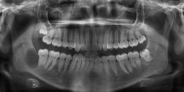
Orthopantomogram (OPG)
The Orthopantomogram (also known as an orthopantomograph, pantomogram, OPG or OPT) is a panoramic single image radiograph of the mandible, maxilla and teeth. It is often encountered in dental practice and occasionally in the emergency department; providing a convenient, inexpensive and rapid way to evaluate the gross anatomy of the jaws and related pathology.
There are multiple indications for this type of radiograph including yet not limited to :
- General dental health evaluation for caries or pulp origin disease
- Trauma assessment for tooth or jaw fractures
- Infection evaluation of sinusitis, periodontitis or periapical abscesses
- Tumor or radicular cyst evaluation
- Temporomandibular joint assessment for disease, fractures or dislocations
- Facial bone disease evaluation
- Foreign body localization
- Salivary stone identification (sialolithiasis)
- Growth and development monitoring of pediatric teeth for location, shape, angle, supernumerary tooth presence and tooth germ absence to prevent or prepare for future aesthetic issues
- Initial and progressive evaluation of orthodontic treatment (note an OPG alone is not usually sufficient for preoperative inspection or prosthesis measurement)
OPG X-ray Can Reveal Dental and Medical Problems Such as:
- Cysts/ growth in the jawbones
- Bone loss/ advanced periodontal disease
- Soft Tissue Screening
- Impacted teeth including wisdom teeth
- Disorders of temporomandibular joint
The Main Advantages Of Panoramic Images/OPGs are:
- Wide coverage of teeth and facial bones including the TMJ (Temporomandibular joint)
- Quick and efficient procedure
- Ease of examination
- Low radiation dose
- Can be used for patients who have restricted mouth opening
- Useful aid for patient education and case presentation
Patient Position
Frequent QnA
During the OPG the arm of the machine will rotate slowly around the head but will not come into contact with the patient. The Radiographer (a technologist trained in medical and dental imaging) will instruct the patient to bite on a small plastic mouthpiece attached to the machine, which keeps the top and bottom teeth separated and helps position the mouth properly in the machine.
For a Lateral Cephalogram you are required to hold still and bite together on the back teeth. Lips should be relaxed. The Radiographer will help ensure that patients are in the correct position before taking the x-ray.