Cone Beam CT (CBCT)
CBCT stands for Cone Beam Computed Tomography.It is a technology used to take three dimensional (3-D) images of your teeth, maxillary sinus, nerve pathways, and bone in the maxillofacial region with a single scan. The CBCT system rotates around the patient in approximately 10-30 seconds, capturing data using a cone-shaped x-ray beam.
CBCT is used when regular two dimensional dental x-rays are not sufficient. With CBCT,clinicians can get highly detailed 3-D views of facial region with 6-15 times lower radiation exposure than a conventional CT scan. This may help with the diagnosis,treatment planning and evaluation of certain conditions.
Why CBCT?
For lesions involving dental and bony hard tissues, CBCT is the imaging modality of choice with higher isotropic voxel resolution, lesser exposure, cost and time taken for scan as compared to conventional CT scan. Hence indicated for treatment planning before implant placement, various Orthodontic, Endodontic, Periodontal, Pedodontics, oral surgery, prosthodontic, ENT, Orthopaedic procedures.
Procedure
The procedure is similar to having an OPG. The examination is painless. You will be asked to sit with your chin resting on a small support shelf to provide accurate positioning. The scanner will move around your head and from this the processer will build and store a three-dimensional image that the radiologist will review and assess.
The three-dimensional image data will also be provided on a disc to the dentist or specialist who referred you, and can be used with other software, for example for implant planning.
- The 3-D data will also be provided on a disc to the dentist who referred you
- Can be used with other software
- Example for implant planning.
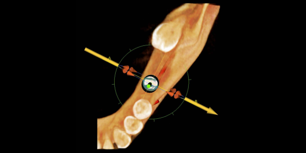
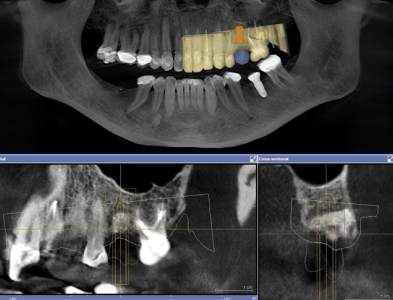
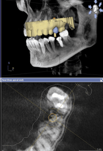
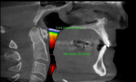
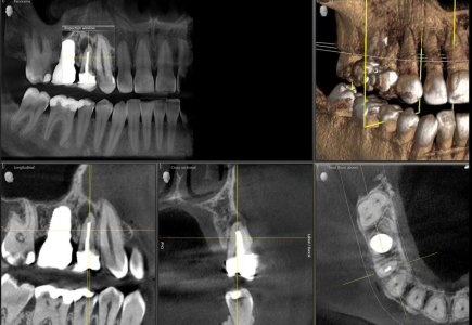
CBCT In Implants
Study consists of cross sections and panoramic projections using Cone Beam CT (CBCT) scanner. It can be of an individual area or the whole mandible or maxilla. CBCT 3D technology is one of the most effective tools available for evaluating implant sites. 3D images can identify potential pathologies and structural abnormalities with unprecedented accuracy.
Cross sectional and panoramic views facilitate various measurements such as: height and width of the implant sites, mandibular edentulous site, a potential implant site near the mental foremen, width of the buccal/lingual ridge and cortical bone density. 3D images highlight the cortical bone thickness, the cancellous bone density, inferior alveolar nerve and the mental foremen location. They also influence the choice of the appropriate implant to be used, its placement, its width and consideration of “die back” from dense cortical bone.
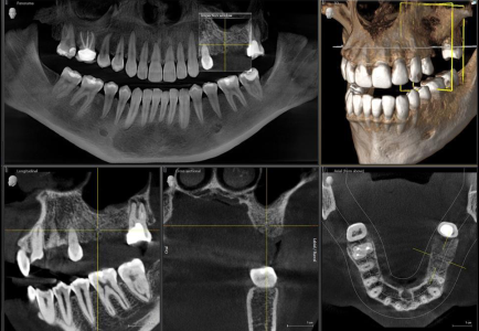
CBCT In Endodontics & Periodontics
In order to perform certain procedures, like treating a fractured tooth, Root canal therapy and caring for the surrounding tissue, endodontic and periodontic specialists require extremely high quality images that will allow them to identify every detail of the treatment area, make an accurate diagnosis, and establish an effective treatment plan. Upon carrying out a thorough examination of the area in question, the user will gain a full appreciation for the device’s less invasive nature and greater suitability.